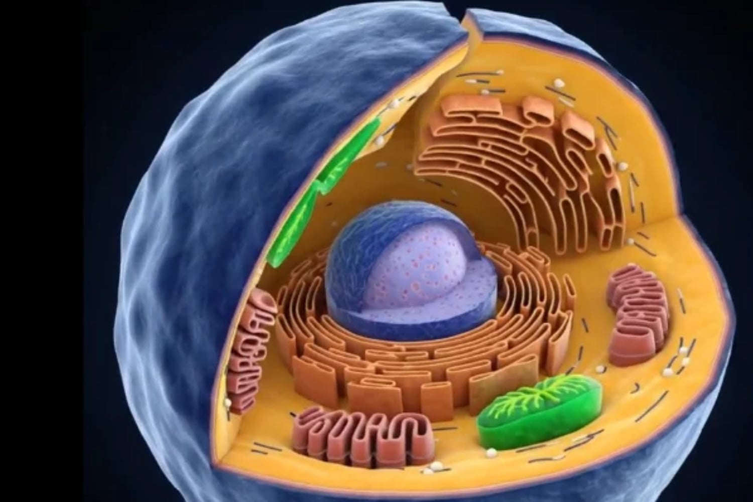
The interior of a cell is filmed in 3D, revealing a world never seen before/Filman el interior de una célula en 3D y revelan un mundo nunca visto
Source
In recent months, several scientific teams have introduced systems capable of observing, in real time and in three dimensions, nanoscale cellular structures that previously could only be seen in fixed (dead) cells or appeared blurred due to movement. Although each team employs a different approach, they share a common goal: to overcome the limitations of traditional microscopy, which either requires high light intensity—thereby damaging the cells—or necessitates a large number of image frames, failing to capture actual movement.
Varios equipos científicos han presentado en los últimos meses sistemas que logran observar, en tiempo real y en tres dimensiones, estructuras celulares de escala nanométrica que antes solo podían verse en células fijadas (muertas) o aparecían borrosas por el movimiento. Aunque cada equipo usa un enfoque distinto, comparten un objetivo: superar las limitaciones de la microscopía tradicional, que exige mucha luz lo que daña las células o muchos fotogramas y no capta el movimiento real.
On one hand, the RO-iSCAT technology approach used by the Australian National University employs a label-free "nanoscopy" technique. The key to this process lies in rotating the angle of the light illuminating the sample and combining images taken at different heights. This eliminates background noise and reveals three-dimensional structures with a light signal ten times stronger—all in real time—without the need for chemical dyes, which can be toxic to cells and even kill them.
Por un lado, el enfoque de la tecnología RO-iSCAT utilizada por la Universidad Nacional de Australia utiliza una técnica de "nanoscopía" sin etiquetas. La clave de este proceso está en rotar el ángulo de la luz que ilumina la muestra y combinar imágenes a distintas alturas. Esto elimina el ruido de fondo y revela estructuras tridimensionales con una señal luminosa diez veces más fuerte en tiempo real, sin necesidad de usar tintes químicos que pueden ser tóxicos para las células y acabar con ellas.
Source
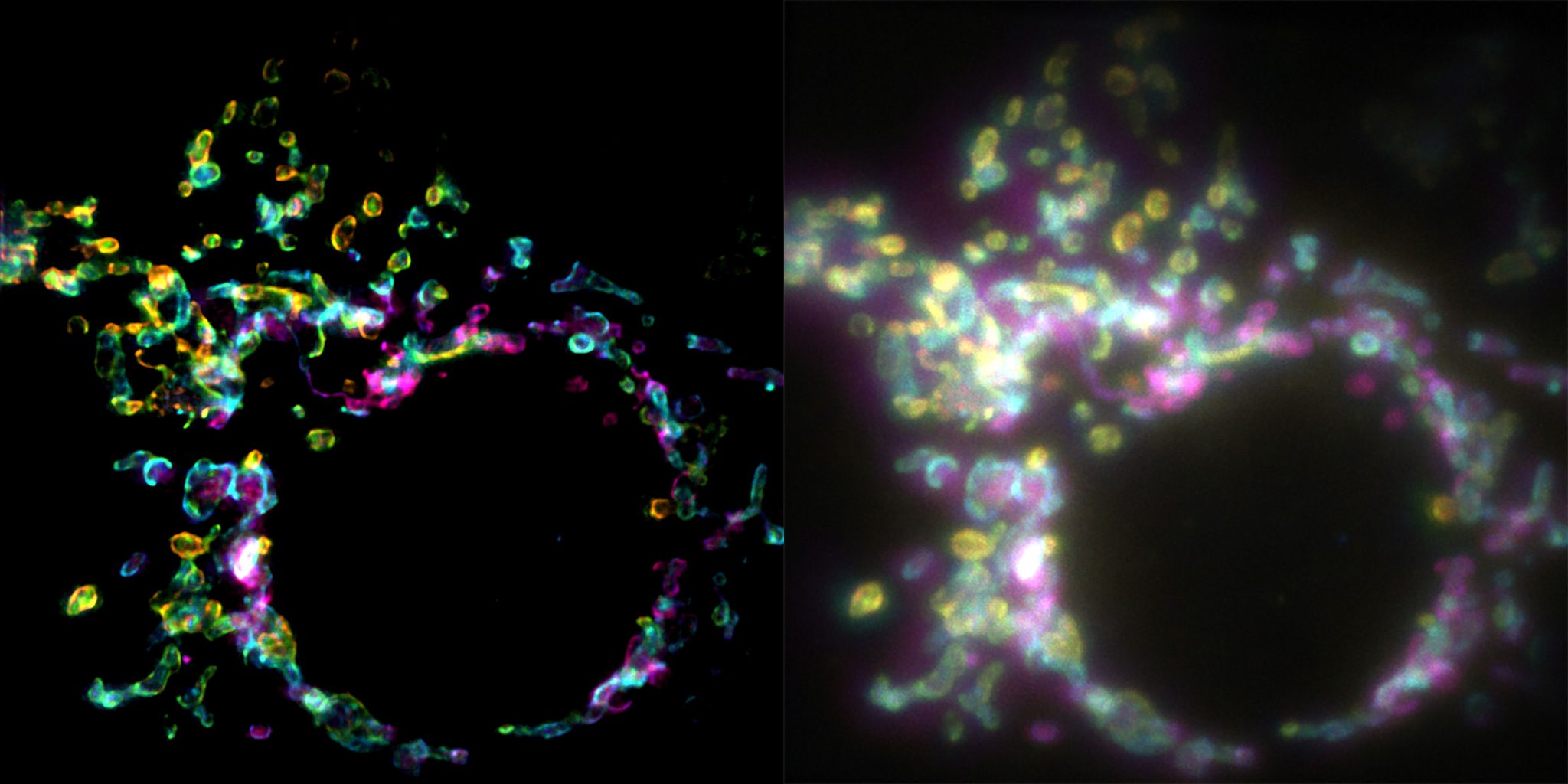
On the other hand, SPIFFI technology (EPFL, Switzerland) is a super-resolution fluorescence technique that, unlike conventional methods, does not require combining hundreds of images captured over time. Instead, it leverages the polarization of the light emitted by fluorescent molecules. By splitting this light into four channels and comparing them, it produces a super-resolution image from a single camera exposure, making it possible to record "videos" of rapid processes, such as the movement of mitochondria.
Por otro lado, la tecnología SPIFFI (EPFL, Suiza), es una técnica de fluorescencia de superresolución que, a diferencia de las convencionales, no necesita combinar cientos de imágenes tomadas en el tiempo. En su lugar, aprovecha la polarización de la luz que emiten las moléculas fluorescentes. Al dividir esa luz en cuatro canales y compararlos, logra una imagen de superresolución con una sola toma de cámara, permitiendo grabar "vídeos" de procesos rápidos como el movimiento de mitocondrias.
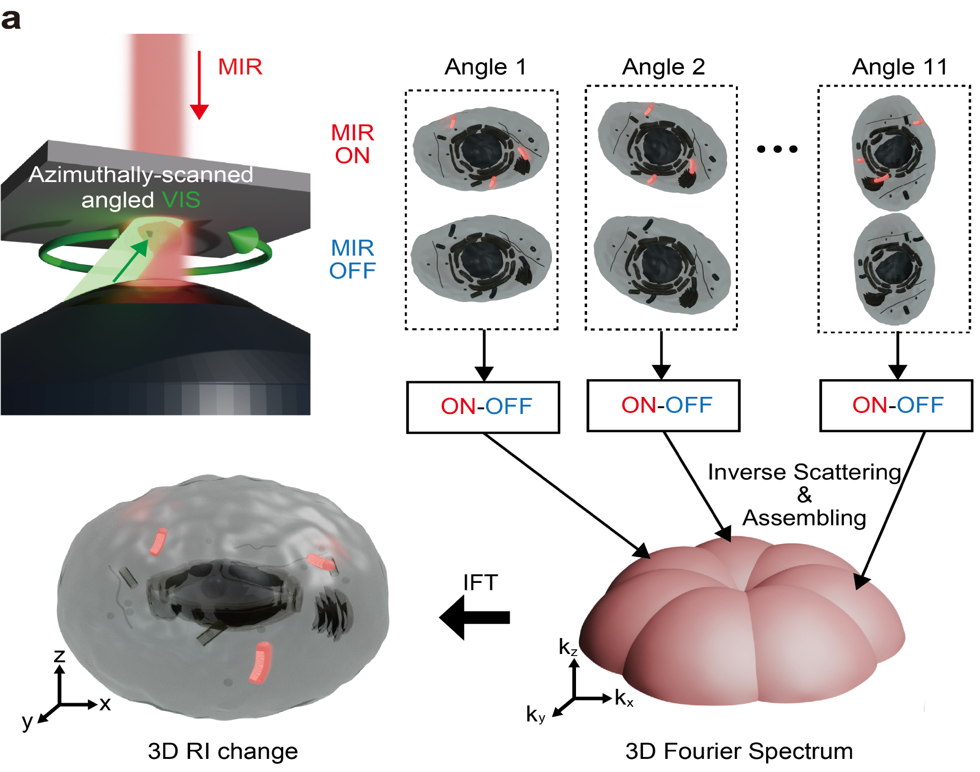
There is also the MIP-ODT technology (University of Tokyo), which uses mid-infrared light to induce vibrations in the chemical bonds of molecules. The resulting heat is detected using visible light. The major breakthrough lies in speed: the system has gone from capturing 0.05 images per second to 19.2 images per second, making it possible to create 3D "videos" of biochemical processes in milliseconds—all without the need for labels. Other approaches exist, such as iISM—developed at Stanford—which combines interferometry with array-based detection to visualize structures as small as 120 nanometers without the use of fluorescent labels, allowing for very long-duration observations without damaging the cell.
También está la tecnología MIP-ODT (Universidad de Tokio) que emplea luz infrarroja media para hacer vibrar los enlaces químicos de las moléculas. El calor resultante se detecta con luz visible. El gran avance es la velocidad, han pasado de capturar 0,05 imágenes por segundo a 19,2 imágenes por segundo, permitiendo crear "vídeos" 3D de procesos bioquímicos en milisegundos, también sin etiquetas. Existen otras aproximaciones como la de iISM desarrollada en Stanford, que combina interferometría y detección por array para ver estructuras de 120 nanómetros sin etiquetas fluorescentes, permitiendo observaciones muy prolongadas sin dañar la célula.
Source
Meanwhile, the STM-FPDT system from Nanjing achieves label-free 3D imaging at a rate of 5 volumes per second, reducing acquisition time by a factor of 4.5. The ability to observe the "secret life" of cells in their natural, dynamic state opens the door to highly significant applications. Using RO-iSCAT, scientists have already observed how pancreatic cancer cells form physical "bridges" with the surrounding connective tissue—a process believed to help tumors grow and resist treatment. It will also enable the study of how viruses (such as HIV) spread between cells via these connections.
Por su parte, el sistema STM-FPDT de Nanjing logra imágenes 3D sin etiquetas a una velocidad de 5 volúmenes por segundo, reduciendo el tiempo de adquisición en 4,5 veces. La capacidad de ver la "vida secreta" de las células en su estado natural y en movimiento abre la puerta a aplicaciones muy relevantes. Con RO-iSCAT, los científicos ya han observado cómo las células de cáncer de páncreas forman "puentes" físicos con el tejido conectivo circundante, un proceso que se cree ayuda a los tumores a crecer y resistir tratamientos. También permitirá estudiar cómo los virus (como el VIH) se propagan entre células a través de estas conexiones.
By observing how living cells respond to a drug over hours or days—without the microscope’s light causing them any damage—it is possible to evaluate the absorption and actual effect of medications within a dynamic environment with much greater precision. Technologies derived from these advances are already being tested for real-time "optical biopsies"—for instance, to distinguish tumor tissue from healthy tissue during surgery, eliminating the need to wait for traditional pathological analysis. The ability to observe the dynamics of "biomolecular condensates" (membrane-less structures that form within cells) could shed light on their malfunction, which is linked to diseases such as Alzheimer’s or Parkinson’s.
Al poder observar durante horas o días cómo las células vivas responden a un medicamento sin que la luz del microscopio las dañe, se puede evaluar con mucha más precisión la absorción y el efecto real de los fármacos en un entorno dinámico. Tecnologías derivadas de estos avances ya se están probando para realizar "biopsias ópticas" en tiempo real, por ejemplo, para distinguir tejido tumoral de tejido sano durante una cirugía sin necesidad de esperar el análisis patológico tradicional. La capacidad de ver la dinámica de los "condensados biomoleculares" (estructuras sin membrana que se forman en las células) podría arrojar luz sobre su mal funcionamiento, que está vinculado a enfermedades como el alzhéimer o el párkinson.
In short, these new technologies are transforming cell biology by allowing us to move from static—and somewhat artificial—"snapshots" to actual "movies" of the internal machinery of life.
En resumen, estas nuevas tecnologías están transformando la biología celular al permitirnos pasar de "fotografías" estáticas y algo artificiales a "películas" reales de la maquinaria interna de la vida.
More information/Más información
https://scitechdaily.com/new-microscope-technology-captures-the-hidden-world-inside-living-cells-like-never-before/
This represents a major breakthrough for cell biology! These new techniques close a huge gap in microscopy by capturing nanoscale structures in real-time 3D without harming live cells. Conventional approaches invariably demanded a trade-off between fine, high-resolution detail and keeping cells alive. Witnessing the dynamic inner world of a living cell revealed in this way will surely speed up our understanding of organelle interactions, disease pathways, and cellular mechanics. What a wonderful step forward!
0.00 SBD,
0.68 STEEM,
0.68 SP
Mirá, eso de rotar el ángulo de la luz para no tener que teñir la célula me parece clave, porque matarla para verla siempre fue una contradicción medio rara. ¿Vos creés que ver ese mundo en movimiento cambia en algo cómo entendemos la vida, o es solo más detalle?
Lo de rotar el ángulo de la luz en RO-iSCAT para eliminar el ruido de fondo sin tintes tóxicos es genial, ahí está la diferencia con la microscopía tradicional. ¿Sabés si ya lo probaron con células vivas en movimiento o solo con muestras fijas?
Lo de RO-iSCAT rotando el ángulo de la luz para eliminar ruido de fondo sin tintes tóxicos es lo que más me convence del artículo. La pregunta es si esa señal diez veces más fuerte se sostiene en muestras vivas durante periodos largos o solo en tomas cortas.
¿Lo de RO-iSCAT sin tintes es lo que más te llamó? xq eso de rotar el ángulo y combinar alturas para eliminar el ruido suena a solución elegante, pero me queda la duda de si sirve igual en células vivas moviéndose o solo en muestras más quietas.
Impresiona que con RO-iSCAT puedan ver estructuras tridimensionales en tiempo real sin tintes que dañen la célula. En mi experiencia, cuando la ciencia logra observar la vida sin destruirla, uno se queda pensando en el cuidado del Creador por lo más pequeño. ¿Ese enfoque sin etiquetas podría aplicarse también a tejidos vivos completos, no solo a células aisladas?